Working Diagnosis:
Chronic left ankle pain secondary to developing osteochondroma with irritation secondary to trauma
Compression of peroneal nerve at fibular head secondary to osteochondroma
Underlying hereditary multiple osteochondromas
Treatment:
After thorough evaluation it was concluded that her fibular pain was secondary to a developing osteochondroma, and aggravated by her prior trauma, as opposed to callus from a healing fracture. The numbness and pain on the lateral aspect of the left ankle was likely secondary to compression of the common peroneal nerve from the osteochondroma. She was initially treated with a home exercises program for motion and strengthening, as well as utilization of the walking boot for long periods of weight bearing activities. She was referred to Orthopedic Oncology and Genetics for the multiple osteochondromas.
Outcome:
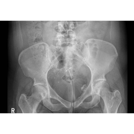
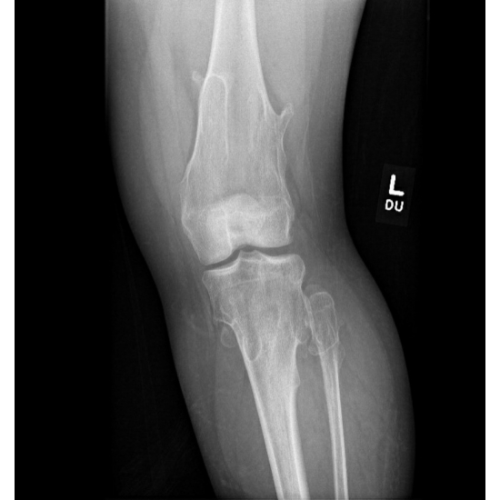
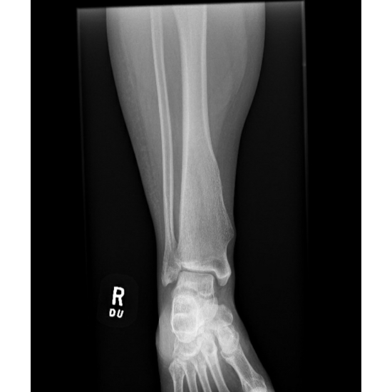
Orthopedic Oncology recommended that she obtain an annual to biannual AP pelvis x-ray to monitor the small osteochondroma projecting from the pelvis as growth of this lesion could not be monitored through physical exam. Case Photo #2 Regarding her remaining osteochondromas, it was recommended to monitor these clinically with further imaging only if she noted an increase in palpable size of lesions or new and/or worsening symptoms. Case Photo #3 , Case Photo #4 For her left ankle, she was taken out of the walking boot completely after her orthopedic oncology appointment.
Gene sequencing for EXT 1 was encouraged after Genetics evaluation to help guide future recommendations related to her health and reproductive decisions.
Author's Comments:
Hereditary multiple osteochondromas (HMO), formerly called multiple hereditary exostoses (MHE), is an autosomal dominant disorder caused by mutations in EXT 1 or EXT 2 tumor suppressor genes that results in a decrease in production of heparin sulfate by chondrocytes. HMO is characterized by multiple osteochondromas which are benign cartilage capped bone tumors which grow from the metaphysis of long bones. Those with EXT 1 mutations have more severe presentations with higher rates of transformation to chondrosarcomas, mo osteochondromas, and more significant limb malalignments.1-3,11
Osteochondromas alone may cause pain, decreased range of motion, local nerve compression (e.g. common peroneal, sciatic), and pathologic fractures. They can also affect growth plates leading to short stature and angular deformities. The most concerning complication of HMO is transformation to a chondrosarcoma.1,11 Patients with HMO have a 2-5% chance of malignant transformation compared to solitary osteochondromas where the risk of malignant transformation is less than 1%.5,11,12 Proximal osteochondromas that involve the pelvis, scapula, proximal femur, and humerus have higher rates of malignant transformation in comparison to distal lesions; the pelvis is the most common location for malignant transformation.7.12 On MRI, the cartilage cap of an osteochondroma is typically greater than 2 cm in children. In adulthood it is typically less than 1 cm. If the cartilage cap in an adult becomes greater than 2 cm this raises concern for malignant transformation.8,9,11 Prognostic factors that correlate with more benign presentation and fewer complications include female sex, involvement of less than 5 sites, and those with EXT 2 mutations.4
The treatment for HMO includes non-operative observation for most patients. Surgical excision of osteochondromas is indicated or considered when pain, deformity, or loss of motion is present or there is evidence or concern for transformation to a chondrosarcoma.7,11 Of note, there is no consensus on criterion for routine imaging surveillance for those with HMO.10 In a study by Wuyts W, Schmale GA, Chansky HA, et al. surveillance recommendations were based on the location of the osteochondroma as more proximal lesions (pelvis, scapula) have higher rates of chondrosarcoma transformation. For proximal lesions, it is suggested that imaging may be done every 2-3 years.10 Research that further investigates a consensus on surveillance criteria based on prognostic factors (sex, age, type of genetic mutation) and/or osteochondroma qualities (quantity, location, size) for HMO would benefit both providers and patients, ensuring appropriate and comprehensive treatment protocols for this potentially debilitating condition.
Editor's Comments:
Osteochondromas are benign bone tumors that develop during childhood or adolescence. They occur due to abnormal growth on the surface of the bone at the growth plate. They are a combination of cartilage and bone. They may occur after a Salter-Harris fracture or surgery. The most common surface areas of bones where these develop are the knee (proximal tibia, distal femur), proximal humerus and proximal femur. While solitary osteochondromas are the most common benign bone tumor, the true incidence is likely unknown as many are asymptomatic.
When patients present for evaluation, symptoms may range from a painless mass at the area of bony growth to bursitis related to friction with overlying soft tissues to neurovascular symptoms if the growth is compressing nearby vessels or nerves. Radiographs demonstrate outgrowths of the bone that can be either sessile or pendunclated. The bony cortex of the lesion is continuous with the bone from which the lesion arises and a radiolucent cartilage cap is often present. In cases where further imaging is needed, CT or MRI can further characterize the lesion.
In asymptomatic individuals, treatment for solitary osteochondromas is observation. When patients are symptomatic, surgical intervention can be considered to resect the lesion. Ideally this should be delayed until skeletal maturity has been reached.
As noted above, if a patient has multiple osteochondromas further work up is recommended due to the higher risk of developing secondary chondrosarcoma in multiple hereditary osteochondroma. Patients with HMO may have more mechanical symptoms due to limb deformities as well pain. Acute onset of pain in patients with HMO should warrant further work up for chondrosarcoma.
References:
1. Pannier S, Legeai-Mallet L. Hereditary multiple exostoses and enchondromatosis. Best Pract Res Clin Rheumatol. 2008;22(1):45-54. doi:10.1016/j.berh.2007.12.004
2. Pei Y, Wang Y, Huang W, Hu B, Huang D, Zhou Y, Su P. Novel mutations of EXT1 and EXT2 genes among families and sporadic cases with multiple exostoses. Genet Test Mol Biomarkers. 2010 Dec;14(6):865-72. doi: 10.1089/gtmb.2010.0040. Epub 2010 Nov 1.
3. J�ger M, Westhoff B, Portier S, Leube B, Hardt K, Royer-Pokora B, Gossheger G, Krauspe R. Clinical outcome and genotype in patients with hereditary multiple exostoses. J Orthop Res. 2007 Dec;25(12):1541-51.
4. Pedrini E, Jennes I, Tremosini M, Milanesi A, Mordenti M, Parra A, Sgariglia F, Zuntini M, Campanacci L, Fabbri N, Pignotti E, Wuyts W, Sangiorgi L. Genotype-phenotype correlation study in 529 patients with multiple hereditary exostoses: identification of "protective" and "risk" factors. J Bone Joint Surg Am. 2011 Dec 21;93(24):2294-302. doi: 10.2106/JBJS.J.00949
5. Tsuda Y, Gregory JJ, Fujiwara T, Abudu S. Secondary chondrosarcoma arising from osteochondroma: outcomes and prognostic factors. Bone Joint J. 2019;101-B(10):1313-1320. doi:10.1302/0301-620X.101B9.BJJ-2019-0190.R1
6. Bov�e JV. Multiple osteochondromas. Orphanet J Rare Dis. 2008;3:3. Published 2008 Feb 13. doi:10.1186/1750-1172-3-3
7. Ahmed AR, Tan TS, Unni KK, Collins MS, Wenger DE, Sim FH. Secondary chondrosarcoma in osteochondroma: report of 107 patients. Clin Orthop Relat Res. 2003;(411):193-206. doi:10.1097/01.blo.0000069888.31220.2b
8. Garrison RC, Unni KK, McLeod RA, Pritchard DJ, Dahlin DC. Chondrosarcoma arising in osteochondroma. Cancer. 1982;49(9):1890-1897. doi:10.1002/1097-0142(19820501)49:93.0.co;2-u
9. Bernard SA, Murphey MD, Flemming DJ, Kransdorf MJ. Improved differentiation of benign osteochondromas from secondary chondrosarcomas with standardized measurement of cartilage cap at CT and MR imaging. Radiology. 2010;255(3):857-865. doi:10.1148/radiol.10082120
10. Wuyts W, Schmale GA, Chansky HA, et al. Hereditary Multiple Osteochondromas. 2000 Aug 3 [Updated 2020 Aug 6]. In: Adam MP, Ardinger HH, Pagon RA, et al., editors. GeneReviews� [Internet]. Seattle (WA): University of Washington, Seattle; 1993-2020. Available from: https://www.ncbi.nlm.nih.gov/books/NBK1235/
11. de Souza AM, Bispo J�nior RZ. Osteochondroma: ignore or investigate?. Rev Bras Ortop. 2014;49(6):555-564. Published 2014 Oct 27. doi:10.1016/j.rboe.2013.10.002
https://www.ncbi.nlm.nih.gov/pmc/articles/PMC4487501/
12. Wittig, James C., et al. �Osteochondroma & Multiple Hereditary Exostosis.� Orthobullets, 8 Dec. 2019, www.orthobullets.com/pathology/8020/osteochondroma-and-multiple-hereditary-exostosis.
Return To The Case Studies List.


